
Technology
The fastest microscope in the world captures electrons down to the attosecond
Electron microscopy has been around for nearly a century, but a record-breaking modern iteration has finally accomplished what physicists have waited decades for: For the first time, a transmission electron microscope captures an electron with such brightness that they can see its individual components. Researchers believe they have opened up a whole new domain of optical science that they now call ‘attomicroscopy’ that will influence the worlds of quantum physics, biology and chemistry.
The breakthrough comes from a team led by experts at the University of Arizona and is detailed in a new study published on August 21 Scientific progress. Mohammed Hassan, associate professor of physics and optical sciences at UA, compares transmission electron microscopes to a smartphone camera.
“If you get the latest version of a smartphone, it comes with a better camera,” Hassan said in an interview accompanying university statement on Wednesday. “… With this microscope, we hope the scientific community can understand the quantum physics behind how an electron behaves and how an electron moves.”
[Related: Winners of the 2023 Nobel Prize in physics measured electrons by the attosecond.]
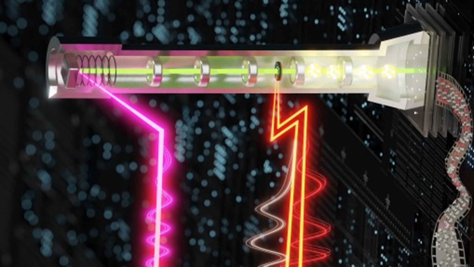
While the original electron microscope arrived in the early 1930s (there is still a controversy about it to this day who invented the very first), scientists have relied on so-called transmission electron microscopes since the 2000s. In these devices, objects are magnified millions of times their scope goes far beyond what light microscopes can achieve. This is because they rely on pulses of electron laser beams fired at a target. From there, high-precision camera sensors and lenses image these atomic particles as they pass through the sample. The changes observed in a subject between these images are called the temporal resolution of a microscope. To increase resolution, researchers have turned to speeding up those laser bursts to attoseconds that last just a trillionth of a second.
But even here the problem is ‘attoseconds’, plural. If physicists ever hoped to capture a single electron frozen in place and detail its incomprehensibly fast subatomic reactions and interactions, they would need a transmission electron microscope capable of firing a single attosecond pulse. To make this a reality, researchers turned to the work pioneered by the 2023 Nobel Prize in Physics winners, who generated the first extreme ultraviolet radiation pulse, also measured in attoseconds. With that foundation, the team finally reached that one attosecond benchmark.
To do this, researchers developed and built a new microscope that splits the laser into a single electron pulse and two pulses of ultra-short light. The first light pulse, a so-called pump pulse, activates the electrons of a sample. A so-called optical gate pulse is then initiated, allowing an infinitesimally small window of time for a one-attosecond electron pulse to then be emitted through the microscope. Once the two ultrashort light pulses are properly synchronized, operators time the electron pulses to help capture atomic events with attosecond-level temporal resolution.
“The improvement of temporal resolution in electron microscopes has long been expected and is the focus of many research groups,” Hassan said on Wednesday. “…For the first time we can see pieces of the electron in motion.”
According to the research summary, the attosecond microscope will enable physicists, optical scientists and other experts to study electron motion in unprecedented detail and “connect this directly to the structural dynamics of matter in real-time and space domains.” This, they say, will hopefully pave the way for “real-life attosecond scientific applications in quantum physics, chemistry and biology.”